Categories
Bergen

Live tissue imaging of protein-protein interactions by multiphoton FLIM-FRET

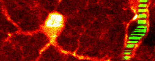
Many signal transduction events in cells are mediated by protein-protein or protein-RNA interactions.  Changes in protein activity are often mediated by a change in protein conformation. Such interactions and conformational changes can be monitored by Förster resonance energy transfer (FRET) between donor and acceptor fluorophores attached to the molecules of interest. The most reliable method for FRET detection in live cells is fluorescence lifetime imaging (FLIM).  FLIM-FRET is based on detecting changes in fluorescence lifetime of the donor fluorophore and is independent of donor and acceptor concentrations in the cell.  

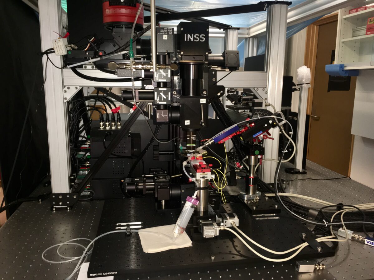
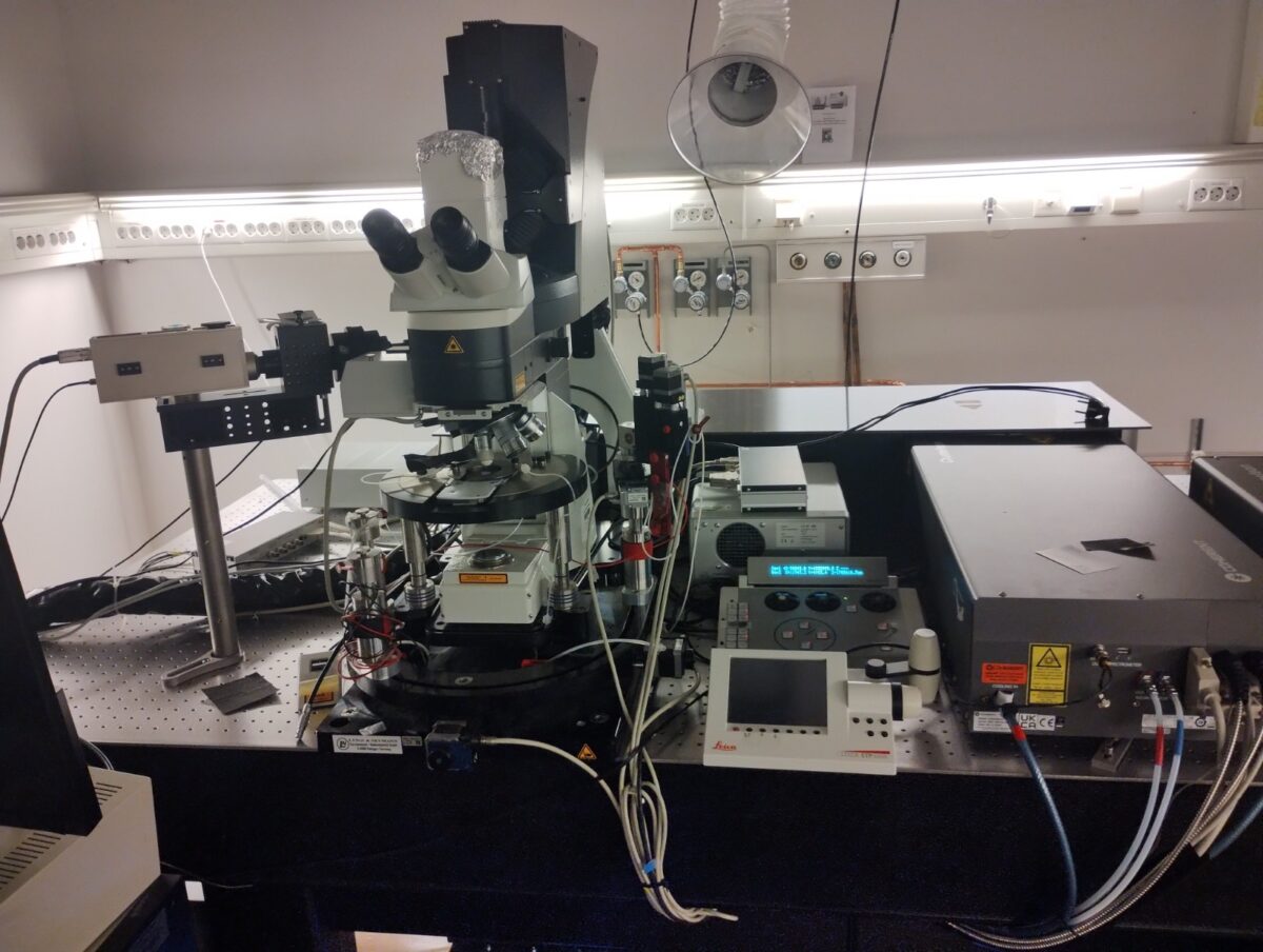
This instrument will allow 2P-based FLIM-FRET imaging in live thick-tissue specimens such as brain tissue slices. The methods require expression of genetically-encoded fluorophores.

The rig is equipped with a Becker & Hickl single-channel FLIM detector and workstation for time-correlated single photon counting (TCSPC). 2P excitation is achieved using a Coherent Chameleon Vision II laser. The FLIM system is integrated with an upright microscope (Leica SP5) for confocal/2P imaging.

Categories
Bergen

3D scanning and uncaging two-photon microscopy

Two-photon (2P) microscopy is an extremely powerful technique that permits visualization and interrogation of complex neural tissue in 3D, with detailed subcellular resolution and minimal phototoxicity. When neurons receive and transform signals arising from hundreds and thousands of synaptic inputs, it is technically extremely challenging to investigate the underlying mechanisms at the necessary spatial and temporal resolution. When 2P microscopy is combined with 2P neurotransmitter uncaging, it becomes possible to precisely activate single and multiple synapses to investigate how dendrites transform synaptic inputs and how neurons and neural microcircuits process information.

This instrument will allow 2P-based structural and functional imaging and neurotransmitter uncaging in live (in vitro) brain tissue slices, combined with multi-electrode patch-clamp recording.

The setup is based on a custom-built 2P microscope (Independent Neuroscience Services, UK) with dual scan paths for resonance-galvo-galvo (RGG) and galvo-galvo scanning, epi- and transfluorescence GaAsP PMTs (Hamamatsu H11706P), fast transimpedance amplifiers (Thorlabs TIA60), and vDAQ acquisition hardware controlled by ScanImage (Vidrio Technologies / MBF Bioscience). Fast 3D scanning and uncaging are implemented by remote focussing units (RFUs) with voice coil actuators (PIMag, Physik Instrumente). 2P imaging and uncaging use a combination of a Mai Tai (eHP DS ; 690-1040 nm tuning range) and InSight X3 (680-1300 nm tuning range) lasers (SpectraPhysics).

Categories
Oslo

Two-photon laser scanning microscopy units

Two-photon laser scanning microscopy is a fluorescence imaging technique that allows fast and minimally invasive imaging of living tissue to a depth of one millimeter at sub-micrometer lateral resolution. Compared to confocal microscopy, 2PLSM offers the advantages of deeper tissue penetration, less photo damage outside the focal region, inherent optical slicing and less chromatic aberration.

The technology allows fast, detailed imaging of calcium signalling at the synaptic level, including measurements of cerebral blood flow (after intravascular injection of fluorescent dye), brain fluid dynamics (after injection of fluorescent tracer in the cerebrospinal fluid), and brain metabolism.

The NORBRAIN node at UiO includes advanced two-photon laser imaging units for:

We offer expertise and imaging technology for a wide range of basic science applications. Depending on project needs and experimental design, access is typically given in a collaboration context. 

Photo: Letten Centre, UiO.

Categories
Trondheim

Two-photon imaging of neural circuit activity in freely-moving rodents

Development of a miniature 2-photon miniscope for large-scale calcium imaging in freely moving mice allows stable simultaneous recording of more than a thousand cells across multiple planes of densely active cortical regions in a wide spectrum of behavioral tasks without impediment of the animal’s behavior.

While the Neuropixels probes increase cell numbers by an order of magnitude, their spatial resolution is as limited as in conventional tetrode-based extracellular recordings. Optical imaging provides a complementary solution to neurophysiological recordings, with high spatial but poorer temporal resolution. The technique uses an indicator molecule such as fluorescent GCaMP6 to monitor changes in intracellular free calcium concentrations optically across wide fields of view (FOV), allowing many hundreds of individual neurons to be followed at near-spike temporal resolution. High spatial resolution has been obtained with two-photon (2P) microscopy but until now this technology was limited to stationary table-top setups, where the animal must be head-fixed under the objective.

Recently, Zong et al. (Cell, 2022) developed a miniaturized two-photon microscope (MINI2P) for fast, high-resolution, multiplane calcium imaging of over 1,000 neurons at a time in freely moving mice. With a microscope weight below 3 g and a highly flexible connection cable, Mini2P allows stable imaging with no impediment of behavior in a variety of assays compared to untethered, unimplanted animals. The improved cell yield is achieved through a optical system design featuring an enlarged field of view (FOV) and a microtunable lens with increased z-scanning range and speed that allows fast and stable imaging of multiple interleaved planes, as well as 3D functional imaging.