Categories
Trondheim

Light-sheet fluorescence microscopy facility

The introduction of genetic tools for labelling have opened the doors to cell-specific connectomic studies, but mapping of brain connectivity still faces labour-intensive processes such as microtomy, histotechnical preparation, and alignment of individual sections into one coherent brain volume to allow for 3D analyses. 3D image volumes captured at microscopic resolution allow for more advanced and efficient analyses. One new approach to obtain such 3D volumes is light sheet microscopy (LSM), a fluorescent technique using optical sectioning of tissue.

Location: NTNU (in collaboration with Leergaard, UIO) 

LSM is based on the concept of illuminating a specimen with a thin sheet of laser light and recording orthogonally the fluorescence emitted from this entire field of view. Originally implemented for small volume samples of transparent tissue, the next generation of instruments enables imaging of chemically cleared mouse brains.The close collaboration between the neuroanatomy cores at NTNU and UIO, part of the Norwegian node of the International Neuroinformatics Coordinating Facility, secures a seamless integration with the established and to be developed digital atlas and analytical pipelines. 

Categories
Oslo

Slidescanning

The equipment delivers high-resolution microscopic brightfield or fluorescence images of histological sections on standard (or large format) slides, with options for acquisitions of entire sections with extended focus range (projection of multiple focal planes) or multiple focal planes.

Equipment

The AxioScan Z1 (Carl Zeiss) allows brightfield or fluorescence digitization of histological sections on standard or large format slides at a resolution of 0.11 micrometers per pixel in ZEN (.czi) format (for ZEN lite Blue from Carl Zeiss) with options for export to TIFF format. The instrument offers extended focus as well as acquisition of multiple focal planes.

Currently the following fluorescence filter sets are installed in AxioScan Z1, detailed description of filter set can be found via links to Zeiss’ website:

Software ‘Zen lite’ from Zeiss can be used for image viewing of ZEN format. It is a free software and please contact us for access.

Photo: Gunnar F. Lothe, Institutt for medisinske basalfag, UiO.

Categories
Oslo

Super resolution microscopy

Super resolution microscopy (SRM) allows optical microscopy with higher resolutions, beyond the light diffraction limit. The NORBRAIN SRM units offers expertise, services, education, and training to enhance biomedical research through super-resolution-based microscopy.

Equipment and software

Zeiss ELYRA PS.1 SIM/STORM/PALM super-resolution microscope

The main advantage of the Zeiss ELYRA PS.1 system is that users can utilize most existing thin-section samples in a fairly turnkey environment with relatively little supervision.

Three distinct imaging modes are available, depending on the sample type:

  1. Structured Illumination Microscopy (SIM) is based on patterns of light being projected in the image plane and subsequent post-processing of these images to form a super resolution image. Most thin flourescent samples with standard dyes work without further processing of samples, resolution ‰250 nm. Several dyes can be used simultaneously, axial (Z) resolution is also improved (for 3D localizations), so that a wide-field image is acquired.
  2. Stochastic Optical Reconstruction Microscopy (STORM) is based on photoswitchable fluorophores to achieve a limited population for sampling. Special chromophores and buffers allow localizations down to ‰20 nm.
  3. Photo Activated Localization Microscopy (PALM) is based on the precise localization of single, sparsely spaced, fluorescent molecules. In order to achieve sparse spacing only a subset of fluorophores are activated at a time, through photoactivation. This requires special fluorophores and longer scan times to build up images. Z-resolution is improved by TIRF, but carries the same limitaions as TIRF. A resolving power of ≈20 nm (≈10X the diffraction limit) is achievable. Genetically encoded protein tags in tissue sections or cells can be localized within ‰20 nm under ideal conditions.

Software: ZEN2011 Sp2

Example images:


Photo: Gunnar F. Lothe, Institutt for medisinske basalfag, UiO.

Categories
Trondheim

Scanners and confocal for advanced functional neuroanatomical studies

Process to achieve advanced knowledge of functional neuroanatomy and systems neuroscience. Studies involve detailed explorations of the anatomical structures and pathways that underlie sensation and perception in each of the sensory modalities. 

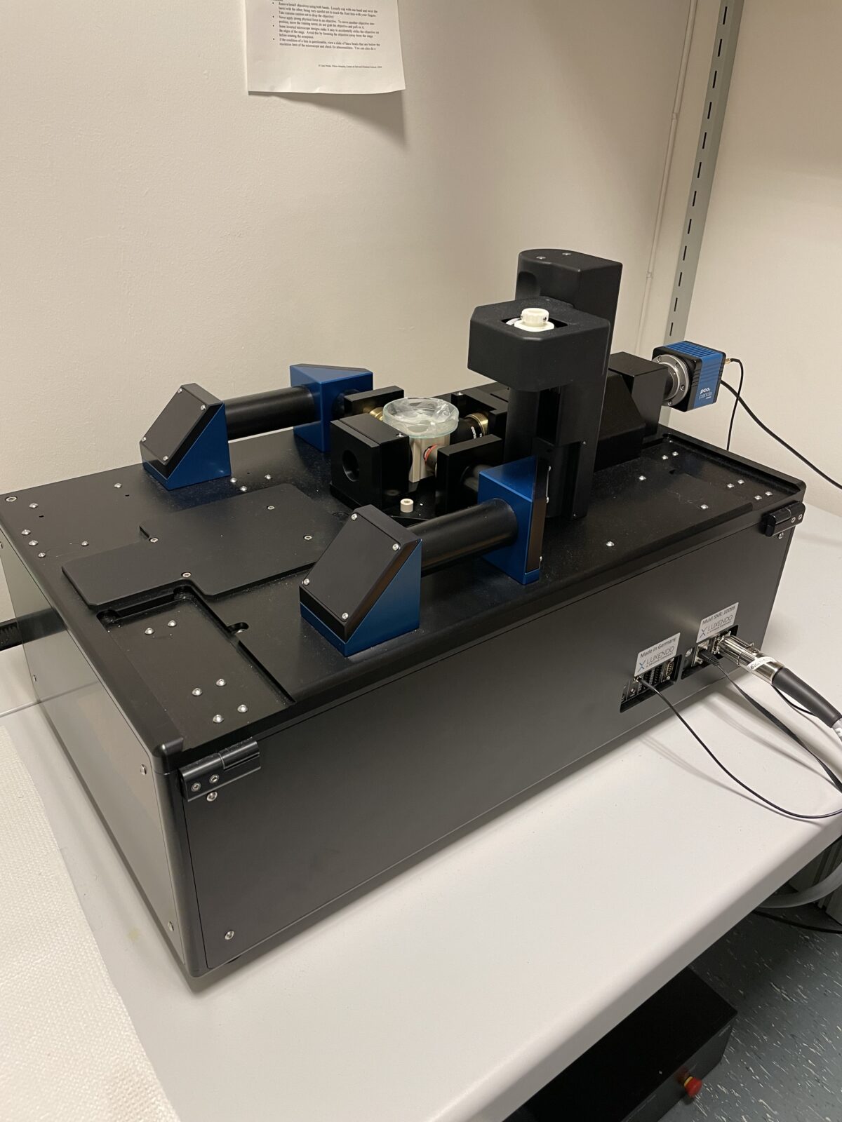
Confocal upright microscope (Zeiss LSM 880) for mounted dry tissue sections and wet specimens 

The confocal laser scanning microscope is an optical imaging technique used to visualize extremely small structures, down to the cellular and sub-cellular size. Its use permits the acquisition of clearer and more detailed pictures when compared with a general widefield microscope, plus the possibility of creating three-dimensional representations of the tissue visualized.  

In our facilities it is possible to use two models of upright Zeiss LSM 880 fluorescence confocal microscope: an AxioImager.Z1 used for mounted dry tissue sections and an AxioExaminer.Z1 for wet specimens. The microscopes are equipped with a range of high quality objectives (10-63x magnification, using air, oil and water as immersion mediums). A varied set of lasers is used as light source, comprising most of the visible spectrum (405, 458, 488, 514, 561, 594 and 633 nm lines). Signal detection is achieved through a spectral detector (34 PMTs, 32 of them being GaAsP detectors). The microscopes are controlled by Zen Black edition software. 

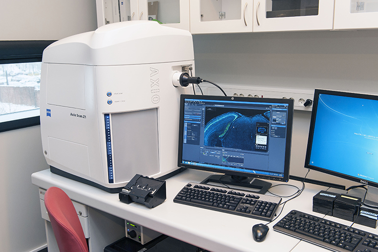
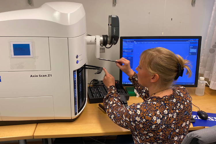
Automated slide scanners 

Two Zeiss AxioScan.Z1 automated slide scanners are available for large-scale widefield digitizing of histological material. Each system fits up to one hundred 26×77 mm microscope slides, it is also  possible to load 52×77 mm slides. In addition to a white lightsource for transmitted light brightfield imaging, the systems are equipped with Zeiss Colibri2 LED sources of different wavelengths for epifluorescence imaging (365, 470. 555, and 625 nm). The two imaging modes employ respectively a 24-bit Hitachi RGB camera and a 16-bit Hamamatsu BW camera. The slide scanners are equipped with high quality air objectives ranging from 5-40X magnification. The systems are controlled by Zen Blue software which also includes options for image processing and analyses such as fluorescence intensity profile.  

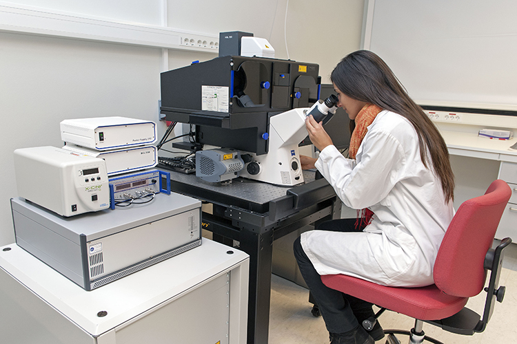
Standalone upright widefield microscopes  

The facility provides one standalone Zeiss AxioImager.M1 upright microscope for checking histological material and taking single, non-tiled images. The stage is manually controlled and can fit 26×77 mm microscope slides. The microscope is equipped with transmitted light brightfield as well as epifluorescent Zeiss Colibri7 LED lightsources (wavelengths 385-630 nm), in addition to air objectives ranging from 1.25-40x. A Zeiss AxioCam MRc camera is fitted on the microscope and is controlled by Zen Blue software.  

For detailed neuroanatomical analyses we provide two upright Zeiss AxioImager microscopes, models M1 and M2. Both microscopes are equipped for transmitted light brightfield as well as epifluorescent imaging and have high quality air and oil objectives ranging from 1.25-100x. The microscopes are also fitted with MicroBrightField CX9000 cameras. The stages are motorized and can fit 26×77 mm microscope slides. The microscopes can also be controlled by MicroBrightfield software installed on the accompanying computers. The StereoInvestigator software is used for unbiased stereology, whereas Neurolucida gives the opportunity to reconstruct neurons or tracing other anatomical structures in 3D. The software may also be used for taking tiled images of smaller areas but not the whole slide. 

Processing and analysis of imaged material 

Three work stations are available for processing and analysis of imaged material. Software installed includes Zeiss Zen Blue Desk (two licenses) for processing .czi images from the AxioScan.Z1 systems, SVI Huygens software for deconvolution of images (one license), and MBF NeuroLucida360 software for 3D neuron/tissue reconstruction and analysis (one license).  

Categories
Bergen

Single-molecule localization microscopy and single-particle tracking (SMLM and SPT)

The Nikon Eclipse Ti2 is a versatile widefield fluorescence microscope optimized for intracellular localization and live-cell tracking of proteins or RNA and detection of cell surface diffusion and membrane trafficking events at the single-particle level. The equipment is suitable for single-molecule localization using photoactivation localization microscopy (PALM), point accumulation in nanoscale topography (PAINT), highly Inclined and laminated optical sheet (HILO) microscopy, total internal reflection fluorescence (TIRF) microscopy, and quantum dot imaging, in addition to common widefield/brightfield microscopy applications. The instrument is suitable for imaging in primary cell cultures, stem cells, cell lines, or similar in vitro preparations.  

For technical specifications see the website of the Molecular Imaging Centre at the University of Bergen.