Categories
Oslo

Microtomy

The microtome park is suited for production of histological sections from fresh or fixed tissues by use of vibratome, freezing microtome or cryostat. The unit includes a hood for dissection and equipment for deparafination and antigen retrieval in formalin fixed, paraffin embedded tissue sections.

Equipment

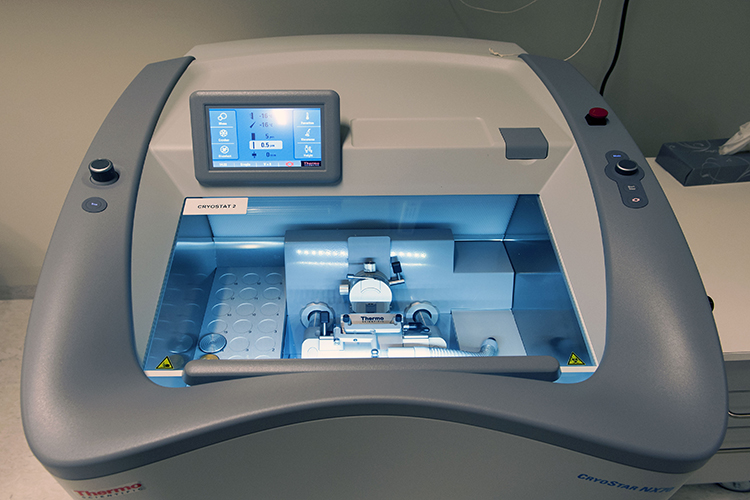
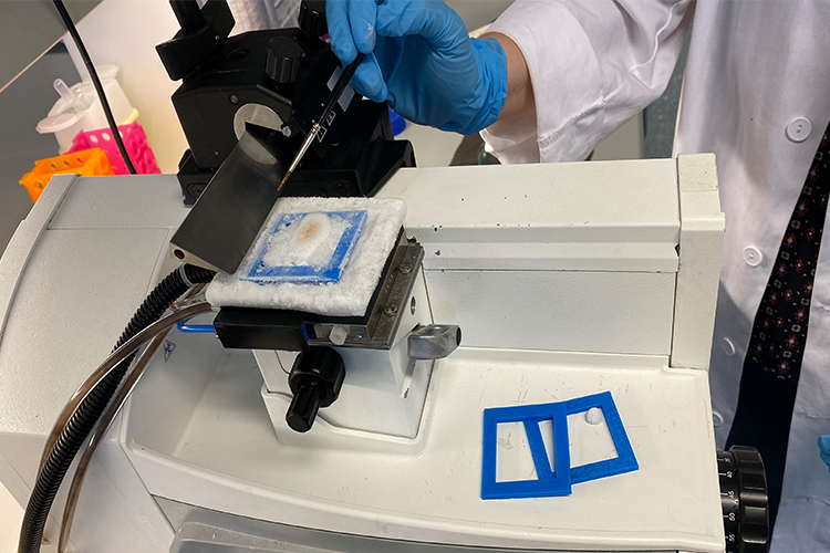
  • Cryostat NX70 (two units) from Thermo Scientific with vacutome, Cold-D disinfection and cooling on knife and objective holder. Suitable for cryosectioning of fixed and fresh tissues at 5 – 20 micrometers.
  • Sliding and freezing microtome MICROM HM 450 (two units). Suited for serial cryosectioning of fixed tissues at 20 – 50 micrometers.
  • Vibratome VT1200S Leica Microsystems with digital camera. Suited for sectioning of relatively thick sections (50-100 µm) of fresh or fixed tissues at room temperature.
  • Dako PT Link is used for deparafination and antigen retrievel from fixed, parafin embedded tissue sections.

Photo: Gunnar F. Lothe, Institutt for medisinske basalfag, UiO.

Categories
Trondheim

Brain slicing and histology

The study of the microanatomy of cells and their formation into tissues and organs as seen through a microscope. Histology  forms the basis of much of our understanding of cell function and disease processes. Our facilities provide equipment for sectioning fresh or fixed tissues in thin slices, as well as resources for visualization of proteins or other components of interest within the tissue. 

Sectioning of tissue 

The facilities offer two Thermo Scientific HM 430  freezing microtomes with external freezing modules. The microtomes are routinely used for sectioning fixed, cryoprotected tissue into slices of 30-50 µm thickness. 

We provide one Leica VT1000S vibratome for sectioning fresh or fixed tissue into slices of 50-400 µm thickness.  

Histological staining procedures 

The facilities include ample space and resources for performing routine histological procedures such as Nissl stain for anatomical delineation and immunohistochemistry for visualization of proteins of interest. We catalogue a wide variety of antibodies as well as multiple fluorescent or chromogenic end products. We have extensive experience in troubleshooting and optimizing immunohistochemical protocols.