Categories
Trondheim

Light-sheet fluorescence microscopy facility

The introduction of genetic tools for labelling have opened the doors to cell-specific connectomic studies, but mapping of brain connectivity still faces labour-intensive processes such as microtomy, histotechnical preparation, and alignment of individual sections into one coherent brain volume to allow for 3D analyses. 3D image volumes captured at microscopic resolution allow for more advanced and efficient analyses. One new approach to obtain such 3D volumes is light sheet microscopy (LSM), a fluorescent technique using optical sectioning of tissue.

Location: NTNU (in collaboration with Leergaard, UIO) 

LSM is based on the concept of illuminating a specimen with a thin sheet of laser light and recording orthogonally the fluorescence emitted from this entire field of view. Originally implemented for small volume samples of transparent tissue, the next generation of instruments enables imaging of chemically cleared mouse brains.The close collaboration between the neuroanatomy cores at NTNU and UIO, part of the Norwegian node of the International Neuroinformatics Coordinating Facility, secures a seamless integration with the established and to be developed digital atlas and analytical pipelines. 

Categories
Trondheim

Viral Vector Core

The Viral Vector Core at Kavli Institute for Systems Neuroscience is a well-equipped facility with a rare expertise to make high quality viruses for research purposes. The Core is a non-profit facility that offers consultation, design and construction of a wide variety of viral vectors to augment scientific research.

Recombinant viruses are highly efficient gene delivery vehicles for introducing molecular tools to difficult-to-transfect primary cells, to create stable cell lines, to transduce tissues in vivo for elucidating underlying biological mechanisms and nowadays for gene therapy applications in clinics.

A combinatorial approach using viral vectors with the vast repertoire of transgenic animals available is a powerful strategy to target specific cells in complex organs such as mammalian brains. 

Adeno-associated virus, Moloney murine leukemia virus, Lenti virus and pseudotyped G-deleted rabies virus are all common transgene-delivery agents, each having distinct characteristics making them suitable for specific research questions.

Depending on the scientific question, one may need viral tools with specific features, demanding novel vector designs.

Recombinant viruses can be custom synthesized on a fee-for service basis, when the customer provides transgene construct for virus production. Vector core can assist in research by designing novel constructs and subsequent production of the viruses, please consult the core facility for the details. 

Categories
Bergen

Live tissue imaging of protein-protein interactions by multiphoton FLIM-FRET

Many signal transduction events in cells are mediated by protein-protein or protein-RNA interactions.  Changes in protein activity are often mediated by a change in protein conformation. Such interactions and conformational changes can be monitored by Förster resonance energy transfer (FRET) between donor and acceptor fluorophores attached to the molecules of interest. The most reliable method for FRET detection in live cells is fluorescence lifetime imaging (FLIM).  FLIM-FRET is based on detecting changes in fluorescence lifetime of the donor fluorophore and is independent of donor and acceptor concentrations in the cell.  

This instrument will allow 2P-based FLIM-FRET imaging in live thick-tissue specimens such as brain tissue slices. The methods require expression of genetically-encoded fluorophores.

The rig is equipped with a Becker & Hickl single-channel FLIM detector and workstation for time-correlated single photon counting (TCSPC). 2P excitation is achieved using a Coherent Chameleon Vision II laser. The FLIM system is integrated with an upright microscope (Leica SP5) for confocal/2P imaging.

Categories
Bergen

3D scanning and uncaging two-photon microscopy

Two-photon (2P) microscopy is an extremely powerful technique that permits visualization and interrogation of complex neural tissue in 3D, with detailed subcellular resolution and minimal phototoxicity. When neurons receive and transform signals arising from hundreds and thousands of synaptic inputs, it is technically extremely challenging to investigate the underlying mechanisms at the necessary spatial and temporal resolution. When 2P microscopy is combined with 2P neurotransmitter uncaging, it becomes possible to precisely activate single and multiple synapses to investigate how dendrites transform synaptic inputs and how neurons and neural microcircuits process information.

This instrument will allow 2P-based structural and functional imaging and neurotransmitter uncaging in live (in vitro) brain tissue slices, combined with multi-electrode patch-clamp recording.

The setup is based on a custom-built 2P microscope (Independent Neuroscience Services, UK) with dual scan paths for resonance-galvo-galvo (RGG) and galvo-galvo scanning, epi- and transfluorescence GaAsP PMTs (Hamamatsu H11706P), fast transimpedance amplifiers (Thorlabs TIA60), and vDAQ acquisition hardware controlled by ScanImage (Vidrio Technologies / MBF Bioscience). Fast 3D scanning and uncaging are implemented by remote focussing units (RFUs) with voice coil actuators (PIMag, Physik Instrumente). 2P imaging and uncaging use a combination of a Mai Tai (eHP DS ; 690-1040 nm tuning range) and InSight X3 (680-1300 nm tuning range) lasers (SpectraPhysics).

Categories
Trondheim

In vitro multi-cell patch clamp electrophysiology

The patch clamp technique is an electrophysiological technique that enables to study the electrical properties of living cells. This method allows to record the ionic currents flowing across the cell membrane (measured in voltage clamp configuration) or changes in membrane potential (voltage, measured in current clamp configuration).  

The electrophysiology rig is optimized for simultaneous patch clamp recordings from multiple neurons (1-4) in slices of brain tissue. Recording multiple cells simultaneously improves the yield of the experiment as well as enables to study properties of communication between cells (synaptic transmission). The rig is equipped with an upright microscope to ensure that suitable cells can be identified and targeted under visual control. Acquisition of electrophysiological data is obtained through a HEKA EPC 10 USB Quadro amplifier, which allows to independently control and record activity from up to four cells simultaneously. The patch clamp rig is further equipped with a laser system containing three different wavelength lasers (375 nm, 473 nm and 594 nm), which enables to combine patch clamp electrophysiological recordings with various optical stimulation techniques. In such experiments, cellular activity can be manipulated by using light-sensitive ligands such as caged neurotransmitters or light-gated ion channels. 

Categories
Oslo

Microtomy

The microtome park is suited for production of histological sections from fresh or fixed tissues by use of vibratome, freezing microtome or cryostat. The unit includes a hood for dissection and equipment for deparafination and antigen retrieval in formalin fixed, paraffin embedded tissue sections.

Equipment

  • Cryostat NX70 (two units) from Thermo Scientific with vacutome, Cold-D disinfection and cooling on knife and objective holder. Suitable for cryosectioning of fixed and fresh tissues at 5 – 20 micrometers.
  • Sliding and freezing microtome MICROM HM 450 (two units). Suited for serial cryosectioning of fixed tissues at 20 – 50 micrometers.
  • Vibratome VT1200S Leica Microsystems with digital camera. Suited for sectioning of relatively thick sections (50-100 µm) of fresh or fixed tissues at room temperature.
  • Dako PT Link is used for deparafination and antigen retrievel from fixed, parafin embedded tissue sections.

Photo: Gunnar F. Lothe, Institutt for medisinske basalfag, UiO.

Categories
Oslo

Slidescanning

The equipment delivers high-resolution microscopic brightfield or fluorescence images of histological sections on standard (or large format) slides, with options for acquisitions of entire sections with extended focus range (projection of multiple focal planes) or multiple focal planes.

Equipment

The AxioScan Z1 (Carl Zeiss) allows brightfield or fluorescence digitization of histological sections on standard or large format slides at a resolution of 0.11 micrometers per pixel in ZEN (.czi) format (for ZEN lite Blue from Carl Zeiss) with options for export to TIFF format. The instrument offers extended focus as well as acquisition of multiple focal planes.

Currently the following fluorescence filter sets are installed in AxioScan Z1, detailed description of filter set can be found via links to Zeiss’ website:

Software ‘Zen lite’ from Zeiss can be used for image viewing of ZEN format. It is a free software and please contact us for access.

Photo: Gunnar F. Lothe, Institutt for medisinske basalfag, UiO.

Categories
Oslo

Super resolution microscopy

Super resolution microscopy (SRM) allows optical microscopy with higher resolutions, beyond the light diffraction limit. The NORBRAIN SRM units offers expertise, services, education, and training to enhance biomedical research through super-resolution-based microscopy.

Equipment and software

Zeiss ELYRA PS.1 SIM/STORM/PALM super-resolution microscope

The main advantage of the Zeiss ELYRA PS.1 system is that users can utilize most existing thin-section samples in a fairly turnkey environment with relatively little supervision.

Three distinct imaging modes are available, depending on the sample type:

  1. Structured Illumination Microscopy (SIM) is based on patterns of light being projected in the image plane and subsequent post-processing of these images to form a super resolution image. Most thin flourescent samples with standard dyes work without further processing of samples, resolution ‰250 nm. Several dyes can be used simultaneously, axial (Z) resolution is also improved (for 3D localizations), so that a wide-field image is acquired.
  2. Stochastic Optical Reconstruction Microscopy (STORM) is based on photoswitchable fluorophores to achieve a limited population for sampling. Special chromophores and buffers allow localizations down to ‰20 nm.
  3. Photo Activated Localization Microscopy (PALM) is based on the precise localization of single, sparsely spaced, fluorescent molecules. In order to achieve sparse spacing only a subset of fluorophores are activated at a time, through photoactivation. This requires special fluorophores and longer scan times to build up images. Z-resolution is improved by TIRF, but carries the same limitaions as TIRF. A resolving power of ≈20 nm (≈10X the diffraction limit) is achievable. Genetically encoded protein tags in tissue sections or cells can be localized within ‰20 nm under ideal conditions.

Software: ZEN2011 Sp2

Example images:


Photo: Gunnar F. Lothe, Institutt for medisinske basalfag, UiO.

Categories
Oslo

Two-photon laser scanning microscopy units

Two-photon laser scanning microscopy is a fluorescence imaging technique that allows fast and minimally invasive imaging of living tissue to a depth of one millimeter at sub-micrometer lateral resolution. Compared to confocal microscopy, 2PLSM offers the advantages of deeper tissue penetration, less photo damage outside the focal region, inherent optical slicing and less chromatic aberration.

The technology allows fast, detailed imaging of calcium signalling at the synaptic level, including measurements of cerebral blood flow (after intravascular injection of fluorescent dye), brain fluid dynamics (after injection of fluorescent tracer in the cerebrospinal fluid), and brain metabolism.

The NORBRAIN node at UiO includes advanced two-photon laser imaging units for:

We offer expertise and imaging technology for a wide range of basic science applications. Depending on project needs and experimental design, access is typically given in a collaboration context. 

Photo: Letten Centre, UiO.

Categories
Trondheim

Very-large-scale Neuropixels silicon probe recordings in freely moving rodents

To understand computation at the neural-circuit level, we must record action potentials simultaneously and accurately from many hundreds of neurons at distributed brain locations in freely moving animals, at a scale very different from that of contemporary studies. 

Conventional tetrode approaches, available in NORBRAIN-1, do not allow for such huge cell samples because each wire has only one recording site, at the tip of the electrode. Silicon probes can have many hundred or thousand sites. NORBRAIN offers access to a new generation of high-density high-site-count Neuropixels silicon probes, in order to prepare the ground for studies, in Norway and elsewhere, that directly address the population mechanisms of neural coding in brain circuits. During 3 years of technology development and testing (2021-23; extended because of the Covid-19 pandemic), we set up several rigs for high density silicon-probe recordings (base stations, headstages, computer equipment) in rodents. 

Neuropixels probes have 960 densely spaced recording sites per electrode shank, from which 384 can be selected. Probes come with 20 µm wide shanks, a 15 mm2 head stage of less than 0.5 g, and on-probe electronic signal buffering and switching circuitry.  

Categories
Trondheim

Two-photon imaging of neural circuit activity in freely-moving rodents

Development of a miniature 2-photon miniscope for large-scale calcium imaging in freely moving mice allows stable simultaneous recording of more than a thousand cells across multiple planes of densely active cortical regions in a wide spectrum of behavioral tasks without impediment of the animal’s behavior.

While the Neuropixels probes increase cell numbers by an order of magnitude, their spatial resolution is as limited as in conventional tetrode-based extracellular recordings. Optical imaging provides a complementary solution to neurophysiological recordings, with high spatial but poorer temporal resolution. The technique uses an indicator molecule such as fluorescent GCaMP6 to monitor changes in intracellular free calcium concentrations optically across wide fields of view (FOV), allowing many hundreds of individual neurons to be followed at near-spike temporal resolution. High spatial resolution has been obtained with two-photon (2P) microscopy but until now this technology was limited to stationary table-top setups, where the animal must be head-fixed under the objective.

Recently, Zong et al. (Cell, 2022) developed a miniaturized two-photon microscope (MINI2P) for fast, high-resolution, multiplane calcium imaging of over 1,000 neurons at a time in freely moving mice. With a microscope weight below 3 g and a highly flexible connection cable, Mini2P allows stable imaging with no impediment of behavior in a variety of assays compared to untethered, unimplanted animals. The improved cell yield is achieved through a optical system design featuring an enlarged field of view (FOV) and a microtunable lens with increased z-scanning range and speed that allows fast and stable imaging of multiple interleaved planes, as well as 3D functional imaging. 

Categories
Trondheim

Brain slicing and histology

The study of the microanatomy of cells and their formation into tissues and organs as seen through a microscope. Histology  forms the basis of much of our understanding of cell function and disease processes. Our facilities provide equipment for sectioning fresh or fixed tissues in thin slices, as well as resources for visualization of proteins or other components of interest within the tissue. 

Sectioning of tissue 

The facilities offer two Thermo Scientific HM 430  freezing microtomes with external freezing modules. The microtomes are routinely used for sectioning fixed, cryoprotected tissue into slices of 30-50 µm thickness. 

We provide one Leica VT1000S vibratome for sectioning fresh or fixed tissue into slices of 50-400 µm thickness.  

Histological staining procedures 

The facilities include ample space and resources for performing routine histological procedures such as Nissl stain for anatomical delineation and immunohistochemistry for visualization of proteins of interest. We catalogue a wide variety of antibodies as well as multiple fluorescent or chromogenic end products. We have extensive experience in troubleshooting and optimizing immunohistochemical protocols.

Categories
Trondheim

7Tesla magnetic-resonance imaging

Norwegian 7T MR Center is a National infrastructure for neuroscience research. The mission is to provide the Norwegian community of neuroscientists the very best tools for high resolution structure-function mapping of the brain.

7 Tesla Magnetic-Resonance (MR) will help to translate knowledge from model preparations in animals to the normal and pathological human brain. While studies of the human brain cannot currently match the spatial and temporal resolution obtained in animal preparations, a clinical 7T MR scanner will, with new technology for reduction of distortions and artefacts, allow us to image the human brain at a very high resolution. This radical improvement in resolution will pave the way for human studies of computation in subareas, layers and columns, which are much closer to the actual computational units of the brain than the global areas accessible with 3T scanners. The anticipated convergence of structural resolution in animal and human studies is likely to link concepts and hypotheses in the two research communities. This is for example critical for studies of entorhinal cortex, which is generally the first brain region to show degeneration in Alzheimer’s disease (AD). We envisage that high-resolution studies of structure, white matter connectivity and grey matter activity patterns on 7T MR scans can be used for subclinical diagnosis and monitoring of disease progression. 

The installed MR-system, a Siemens MAGNETOM Terra System, is equipped with:  

  • Dual Mode (Clinical and Research).  
  • 8 Channel RF-transmit chain in research mode  
  • 80/200 Gradient System.  
  • 32RX/1TX head coil (clinical mode)  
  • 32RX/8TX head coil (research mode only)  
  • 28RX/1TX knee coil (clinical mode)  
  • 13C and 31P loop coils  
  • Multinuclear option  
  • fMRI equipment  


Photo: Siemens Healthineers.  

Categories
Trondheim

Scanners and confocal for advanced functional neuroanatomical studies

Process to achieve advanced knowledge of functional neuroanatomy and systems neuroscience. Studies involve detailed explorations of the anatomical structures and pathways that underlie sensation and perception in each of the sensory modalities. 

Confocal upright microscope (Zeiss LSM 880) for mounted dry tissue sections and wet specimens 

The confocal laser scanning microscope is an optical imaging technique used to visualize extremely small structures, down to the cellular and sub-cellular size. Its use permits the acquisition of clearer and more detailed pictures when compared with a general widefield microscope, plus the possibility of creating three-dimensional representations of the tissue visualized.  

In our facilities it is possible to use two models of upright Zeiss LSM 880 fluorescence confocal microscope: an AxioImager.Z1 used for mounted dry tissue sections and an AxioExaminer.Z1 for wet specimens. The microscopes are equipped with a range of high quality objectives (10-63x magnification, using air, oil and water as immersion mediums). A varied set of lasers is used as light source, comprising most of the visible spectrum (405, 458, 488, 514, 561, 594 and 633 nm lines). Signal detection is achieved through a spectral detector (34 PMTs, 32 of them being GaAsP detectors). The microscopes are controlled by Zen Black edition software. 

Automated slide scanners 

Two Zeiss AxioScan.Z1 automated slide scanners are available for large-scale widefield digitizing of histological material. Each system fits up to one hundred 26×77 mm microscope slides, it is also  possible to load 52×77 mm slides. In addition to a white lightsource for transmitted light brightfield imaging, the systems are equipped with Zeiss Colibri2 LED sources of different wavelengths for epifluorescence imaging (365, 470. 555, and 625 nm). The two imaging modes employ respectively a 24-bit Hitachi RGB camera and a 16-bit Hamamatsu BW camera. The slide scanners are equipped with high quality air objectives ranging from 5-40X magnification. The systems are controlled by Zen Blue software which also includes options for image processing and analyses such as fluorescence intensity profile.  

Standalone upright widefield microscopes  

The facility provides one standalone Zeiss AxioImager.M1 upright microscope for checking histological material and taking single, non-tiled images. The stage is manually controlled and can fit 26×77 mm microscope slides. The microscope is equipped with transmitted light brightfield as well as epifluorescent Zeiss Colibri7 LED lightsources (wavelengths 385-630 nm), in addition to air objectives ranging from 1.25-40x. A Zeiss AxioCam MRc camera is fitted on the microscope and is controlled by Zen Blue software.  

For detailed neuroanatomical analyses we provide two upright Zeiss AxioImager microscopes, models M1 and M2. Both microscopes are equipped for transmitted light brightfield as well as epifluorescent imaging and have high quality air and oil objectives ranging from 1.25-100x. The microscopes are also fitted with MicroBrightField CX9000 cameras. The stages are motorized and can fit 26×77 mm microscope slides. The microscopes can also be controlled by MicroBrightfield software installed on the accompanying computers. The StereoInvestigator software is used for unbiased stereology, whereas Neurolucida gives the opportunity to reconstruct neurons or tracing other anatomical structures in 3D. The software may also be used for taking tiled images of smaller areas but not the whole slide. 

Processing and analysis of imaged material 

Three work stations are available for processing and analysis of imaged material. Software installed includes Zeiss Zen Blue Desk (two licenses) for processing .czi images from the AxioScan.Z1 systems, SVI Huygens software for deconvolution of images (one license), and MBF NeuroLucida360 software for 3D neuron/tissue reconstruction and analysis (one license).  

Categories
Bergen

Single-molecule localization microscopy and single-particle tracking (SMLM and SPT)

The Nikon Eclipse Ti2 is a versatile widefield fluorescence microscope optimized for intracellular localization and live-cell tracking of proteins or RNA and detection of cell surface diffusion and membrane trafficking events at the single-particle level. The equipment is suitable for single-molecule localization using photoactivation localization microscopy (PALM), point accumulation in nanoscale topography (PAINT), highly Inclined and laminated optical sheet (HILO) microscopy, total internal reflection fluorescence (TIRF) microscopy, and quantum dot imaging, in addition to common widefield/brightfield microscopy applications. The instrument is suitable for imaging in primary cell cultures, stem cells, cell lines, or similar in vitro preparations.  

For technical specifications see the website of the Molecular Imaging Centre at the University of Bergen.

Categories
Oslo

Mass spectrometry

The NORBRAIN mass spectrometry unit delivers high sensitivity for the quantitative analysis of small molecules, peptides, toxins and drugs. We provide expertise, services, education, and training to enhance biomedical research through mass spectrometry-based technologies.

Equipment and software

Thermo Scientific Q Exactive Hybrid Quadrupole-Orbitrap Mass Spectrometer

High resolution and accurate mass (HR/AM) analysis on the NORBRAIN Q‐Exactive mass spectrometer presents a unique configuration: the combination of the orbitrap mass analyzer with a quadrupole mass filter for precursor ion mass selection enables new quantitative methods based on HR/AM measurements, including targeted analysis in MS mode (single ion monitoring, SIM) and in MS/MS mode (parallel reaction monitoring, PRM).

The ability of the quadrupole to select a restricted m/z range allows overcoming the dynamic range limitations associated with trapping devices like the Orbitrap XL. Therefore high end quantitative and qualitative analysis in the area of in drug discovery, proteomics, environmental and food safety, clinical research and forensic toxicology can be offered. For example a proteomic discovery analysis on the Q-Exactive allows the detection of several thousand proteins in one run (180 min gradient).

Thermo Scientific TSQ Vantage Triple Stage Quadrupole Mass Spectrometer

Delivers high sensitivity for the quantitative analysis of small molecules, peptides, toxins and drugs. It is coupled online to a Dionex LC-system (Thermo-Fisher Scientific) and offers selected-reaction monitoring of peptide for protein quantification with an extended mass range of up to 4000 m/z.

Both complementary mass spectrometry systems (TSQ Vantage and Q-Exactive) therefore allow to develop high throughput biomarker analyses (TSQ Vantage) based on the suggested biomarker candidates from previous Q-Exactive MS analyses.

Software

  • Database search tools: Sequest (Proteome Discoverer 1.4)  for tandem mass spectra interpretation
  • MaxQuant/ SIEVE for relative quantification
  • In-house software tools (SAPA- and peptide mass tool) for protein peptide analyses

Services

We advise on epiproteomics, protein and lipid profile characterization as well as metabolomics, small molecule analysis, and analysis of post-translational modifications. We also offer services related to quantitative protein expression profiling (SILAC, N15 labelling, LFQ=label free quantification), and consult on experimental design and proteomic analysis options.

  • Protein analyses offered include in-gel or in-solution protease digestion, chromatographic separation and tandem mass spectrometric analysis of the generated peptides, and interpretation of MS/MS data using software like Sequest, Mascot, and MaxQuant.
  • The facility also assists especially in applications for the isolation (enrichment strategies like lectin chromatography), detection and characterization of post-translationally modified peptides (e.g. phosphorylation, N- and O-linked glycosylation, oxidation, ubiquitination, cysteine modifications, acetylation/ methylation (+ di- and tri-methylation) and O-GlcNAc modification). Sites of modification are verified by manual inspection of the data.

Please consult facility staff for projects which implements specialized approaches related to proteomic PTM analysis.


Photo: Gunnar F. Lothe, Institutt for medisinske basalfag, UiO.

Categories
Trondheim

Surgery facilities

Surgery facilities are available for users planning to perform experiments with instruments that are part of the NORBRAIN facility (tetode recordings, Neuropixels recordings, 2P microscope imaging, etc).  

Our surgery facilities are equipped with setup for anesthesia and surgery for intracranial delivery of substances or implantation of devices, in mice or rats. Necessary equipment for supportive therapy and postoperative care are also available, along with consumable materials and storage possibilities for medicines and test substances. 


Storage of medicines and test substances 

Ventilated cabinets are available for storage at room temperature, and fridges and freezers are available for products that need to be stored at cold temperatures.  

Instrument sterilization 

Surgical instruments must be sterilized before they can be used in surgeries. We have equipment for both dry heat sterilization and steam autoclaving. 

Specifications: 

Dry heat sterilization is done using a Termaks drying oven with adjustable temperatures and a display to control the temperature.  

For steam autoclaving we have a Tomy high-pressure steam sterilizer. 

Alternatively, glass bead sterilizers may be used to re-sterilize instruments.  

Anesthesia 

Animals must be anesthetized during surgeries. We have setup for gas anesthesia, which is easy to adjust to achieve suitable depth and a balanced anesthesia. 

Specifications: 

All surgery tables are set up with a vaporizer for either isoflurane or sevoflurane, with the possibility to use medical air and oxygen as the carrier gas. Medical air is delivered from a central system in the building. Oxygen is delivered from oxygen concentrators located in the surgery rooms. The gas anesthesia can be delivered to an induction chamber for induction of anesthesia, to a coaxial mask for maintenance of anesthesia, or to the stereotaxic mask for maintenance during the surgical procedure. The adjustment of the delivery pathway can easily be done by the user. To limit the exposure of the user, the surgery tables are down-drafted and we have an adjustable hood above the table. 

Stereotaxic setup 

The stereotaxic setup is especially designed to do find the correct area of the brain, with the use of precision instruments and coordinates based on anatomical landmarks. This gives the possibility to reach the exact area you are interested in. 

Specifications: 

The surgery tables are equipped with stereotaxic frames from Kopf Instruments, either a model 1430-B “U” frame for rats, or a model 900 “U” frame for mice. We have two main types of manipulators that can be used with the frame: 

  • Model 1760 Micro Manipulator (10 micron resolution): X, Z adjustment – Metric Vernier scale 80 mm travel, calibrated dial – 10 micron increments, 1.0 mm of travel per revolution. Y adjustment – Manual adjustment 100 mm each side of zero (A/P bar) 0.1 mm Vernier scale. 
  • Model 1460 Electrode Manipulator (100 micron resolution): X, Z adjustment – 80 mm travel calibrated 0.1 vernier scale (100 micron increments), 3.0 mm advance per revolution. Y adjustment – Manual adjustment 100 mm each side of zero (A/P bar) 0.1 mm Vernier scale 

The manipulators are used to locate the correct area, or as holders for drill, implants, syringes or pumps. 

Drill 

Specifications: 

Micro motor handpiece drills from Foredom Electric are available on the surgery tables. The drills are small and flexible, they have speed control and can be adjusted to either manual control or foot-pad control. The drills can be integrated with the manipulators on the stereotaxic setup. 

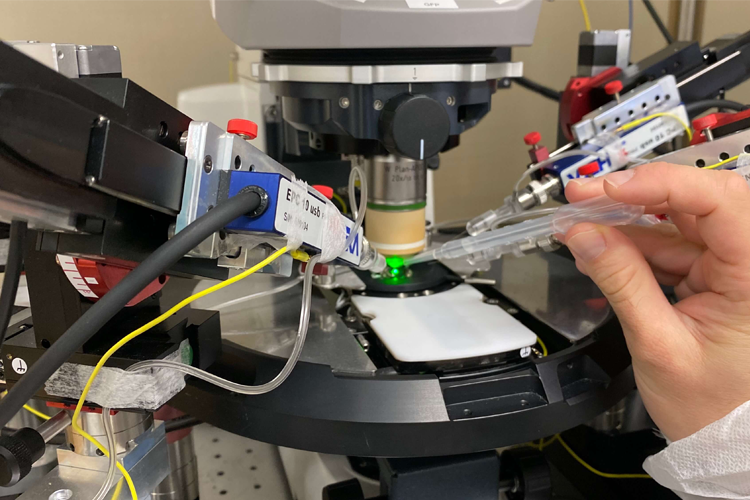
Systems for micro-injection 

When the surgery is done to deliver a small volume of a substance to a localized area in the brain, it is necessary to use special equipment to do this with high precision. 

Specifications: 

Several systems for delivery of substances in the microliter or nanoliter scale are available.  

  • Hamilton syringes have ultrafine needles and can deliver substance in the nanoliter scale. From Hamilton Company 
  • Nanoliter 2010 micro-injection system, which can be combined with a Micro 4 microsyringe pump controller, from World Precision Instruments 
  • Nanoject III programmable nanoliter injector, from Drummond Scientific Company 

Light source 

It is important to have sufficient light when doing surgeries. On the surgery tables we have lamps with flexible tubes so that the user can direct the light to the area where the light is needed. 

Specifications: 

The light source is LED connected to fiberoptic tubes. The user can adjust the direction of the light, and also the light strength, through a dimmer. 

Heat support 

Providing heat support during the surgery is important to maintain the body temperature of the rodents. 

Specifications:  

The surgery tables are equipped with an electric heat pad that is located on top of the platform where the animal is placed during surgeries. The heat pad is connected to a temperature controller with a display to monitor.  

Fluid support 

To prevent dehydration, fluids are given to the animals. Fluids are heated to body temperature.  

Specifications: 

A water bath with adjustable temperatures facilitates the preparation of body-temperature fluids which are given as fluid therapy to the animals during surgeries.  
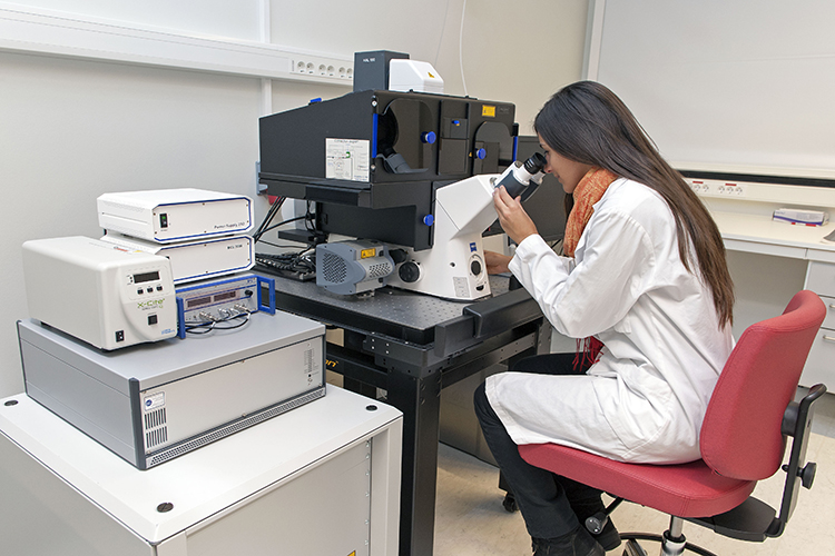
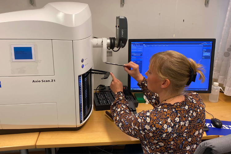
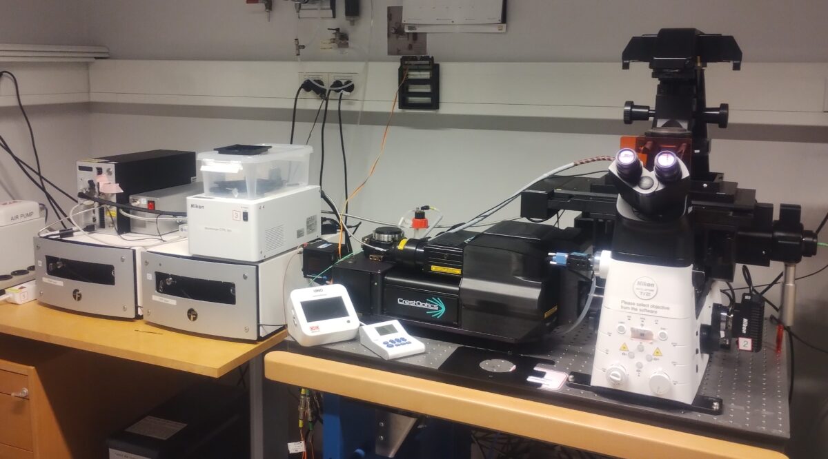
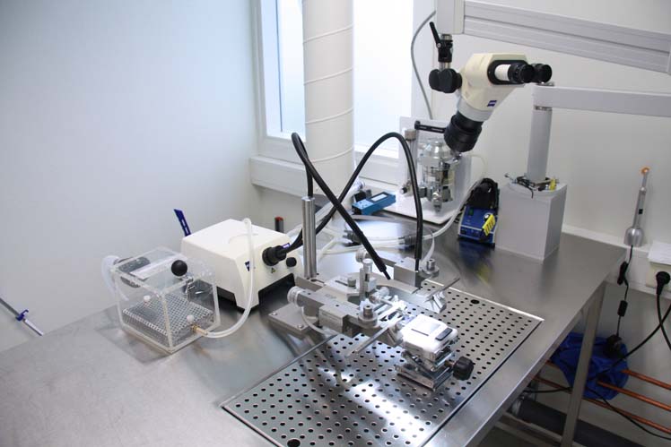
Stereo microscope 

In order to get a good overview of the surgical field, the microscope gives a nice opportunity to see the area and make necessary adjustments. 

Specifications: 

We have stereo microscopes from Nikon and Zeiss, with adjustable focus and zoom. Some of the microscopes are connected to a camera that transfers the view to a screen, so that observers can follow the surgery. 
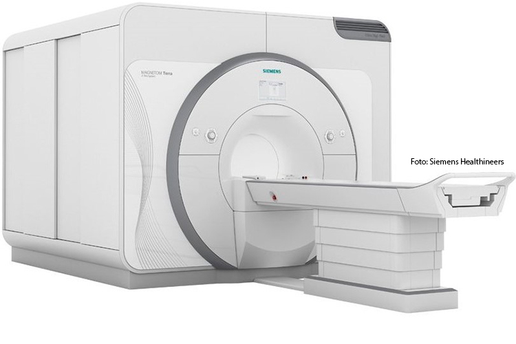
Ultrasound imaging 

One room has a high-frequency ultrasound imaging system that gives high-resolution images in space and time, of small anatomical structures. This gives the possibility to do image-guided procedures, for example. 

Specifications: 

The ultrasound scanner is a Vevo 1100 from Visualsonics, with a MS550S transducer (20-40 mHz and 16 mm penetration), an integrated rail system for positioning the animal and an image-guided injection system. A physiology monitor helps to follow the vital body functions during the procedure. The setup is connected to gas anesthesia. Available functionalities on the ultrasound scanner are M mode, B mode, colour doppler, pulse wave doppler, needle guide, and storage and post-processing of images and videos. 

Post-operative recovery chamber 

The surgical facilities are equipped with recovery chambers where animals can wake up from surgeries. The recovery chambers give the animals a warm and calm environment for the first hours after surgery. 

Specifications: 

The recovery chamber is a Maxi-Thermacage from Datesand. The chamber allows for precise temperature adjustment and control through a display. The temperature is evenly distributed inside the chamber. 

Other laboratory equipment 

The surgical facilities are also equipped with general laboratory tools such as shakers (for example MS3 basic, IKA), micro-sentrifuges (Mini Star, VWR), an ice cube machine (Porkka), and a water purification system (Direct-Q, Merck Milipore). 

Please note: Participating during animal experiments requires that the following conditions are met: 

  • People need to have completed the education and training in laboratory animal science as required by the national competent authority (The Norwegian Food Safety Authorities) equivalent to, at minimum, function (a) (persons who perform procedures on animals) according to the EU commission’s framework document for education and training. The local person responsible for animal care will need to evaluate the documents/certificates of the completed education and training. 
  • The experiments must be approved by the Food Safety Authorities, and the local animal welfare body must be oriented about the experiment in advance. The animal welfare body can set conditions for the experiment, based on local routines and guidelines 
  • People participating in the experiment must have been introduced to local routines regarding, for instance, health and safety, and use of the animal facility 
  • People must have been trained in the correct use of the equipment